Title: Phases of The Cell Life Cycle in Root Tips
Purpose: To observe and determine the phases of the cell life cycle by identifying stages of mitosis in a root tip.
Introduction: Cells at the tip of a growing plant are constantly dividing, allowing the root to grow. In Mitosis, a cell doubles it’s chromosomes and then divides into two identical copies of the original cell. These cells divide independently, thus the cells in the root tip are at different stages of cell division. These stages include interphase, the pre-phase where a cell is just a dark mass as the cell doubles it’s unorganized chromosomes and prepares for division. Prophase, where the chromosomes are visible in a microscope as they condense. Centrioles move to opposite sides of the cell. In Metaphase, the spindle fibers move the chromosomes to the equator of the cell and centrioles are at opposite ends. Next comes Anaphase where the sister chromatids are pulled apart to opposite poles. Finally in Telophase, two daughter cells form and each have a full set of chromosomes as well as a nucleus.
Materials:
- Onion root-tip slides
- Microscope (400x)
Methods:
- Set onion root-tip slide on the set-up microscope stage
- Focus the microscope to clearly view the onion root under the 40x lens, once viewed, change the lens to 400x and focus finely until the cells are clear
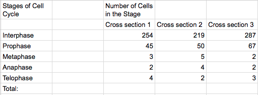
- Count vertically the longest line of cells, then count horizontally the longest row of cells. Multiply these two numbers, the result is the total number of cells in the stage.
- Identify the cells in the phases of Telophase, Anaphase, Metaphase and Prophase, in this order. Record the number of each phase in a table
- Calculate the total number of cells in each phase of the previous step, then subtract that number from the total number of cells in the stage. The result is the number of cells in Interphase, record in the table.
- Looking at two more onion root-tips, repeat steps 2-5 and record.
Data Table (Personal)
Data Table (Class Averages)

Conclusion
- The majority of the cells were in Interphase
- Percentages in each stage
- Interphase: 81%
- Prophase: 12.43%
- Metaphase: 1.65%
- Anaphase: 1.37%
- Telophase: 2.03%
- The evidence that shows that mitosis is a continuous process is that each phase is hard to differentiate which showcases how the process is a flow and not definitive.
- In each cell observed, there are 4X chromosomes being that Interphase doubles the original 2X chromosomes.
- After Meiosis, each sex cell would have 1X chromosome being that each daughter cell (2X) divides in half.
- In the zygotes produced, there would be 2X chromosomes.
Based on the data recorded from the class, the cells are in Interphase 81% of the time which is completely reasonable being that the cells need to function as opposed to just reproducing.

Quinlyn, I love seeing real cells under the microscope. Just wait until next semester when we focus on Kingdom Protista. Lots of live critters will be examined!
LikeLiked by 1 person
I lot of information from an onion root!
LikeLiked by 1 person
I know lab reports are no ones favorite, but we do them at howard so get used to it!!!
LikeLiked by 1 person